Weasis: Free DICOM viewer
A free/libre DICOM viewer
Weasis is a multipurpose standalone and web-based DICOM viewer designed with a highly modular architecture. It is a very popular clinical viewer used in healthcare by hospitals, health networks, multicenter research trials, and by patients.
As free/libre and open source software (FLOSS), Weasis is cross-platform, multi-language, and supports flexible integration with PACS, RIS, HIS, or EHR systems. The viewer is compatible with Windows, Linux, and macOS across various processor architectures (see the available download packages).
Leveraging the OpenCV library, Weasis delivers high-performance and high-quality medical imaging renderings.
From version 4 onwards, Weasis features a responsive user interface aligned with operating system options, offering an enhanced experience on high-resolution screens.
-
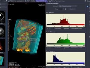 Color Histograms
-
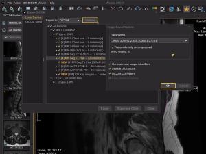 Exporting DICOM
-
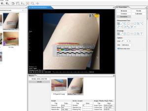
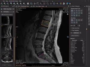 Measurement tools
-
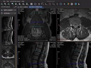
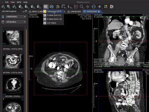 Orthogonal Multiplanar Reconstruction
-
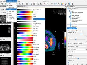 Pseudo-color
-
 User Interface Language
Weasis has been designed to meet the evolving needs of clinical information systems in medical imaging. Its features focus on providing web-based access to radiological images, supporting a wide range of DICOM types, and offering multimedia capabilities.
Key Viewer Features (see also Tutorials)
-
Data type support
- Weasis provides a highly detailed implementation of the DICOM standard, enabling effortless display and interaction with most types of DICOM files.
- Display most DICOM files including multi-frame, enhanced, MPEG-2, MPEG-4, MIME Encapsulation, DOC, SR, PR, KOS, SEG, AU, RT, and ECG
- Display DICOM image containing float or double data (Parametric Map)
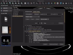
- Import DICOM files with DICOM Query/Retrieve (C-GET, C-MOVE and WADO-URI) and DICOMWeb (QUERY and RETRIEVE)
- Import and export DICOM CD/DVD with DICOMDIR
- Import and export DICOM ZIP files
- Viewer for common image formats (TIFF, BMP, GIF, JPEG, PNG, RAS, HDR, and PNM)
-
Exporting data
- Export DICOM files locally with several options (DICOMDIR, ZIP, ISO image file with Weasis, TIFF, JPEG, PNG…)
- Send DICOM files to a remote PACS or DICOMWeb server (C-STORE or STOW-RS)
- Save measurements and annotations in DICOM Presentation States or XML file
-
Viewing and image rendering
- Support of several screens with different calibration, support of HiDPI (High Dots Per Inch) monitors, full-screen mode
- Image manipulation with mouse buttons (pan, zoom, windowing, rotation, scroll, crosshair)
- Support of DICOM Modality LUTs, VOI LUTs, LUT Shapes, and Presentation LUTs (even non-linear)
- Apply DICOM Presentation States (GSPS) and display graphics as overlays
- Support DICOM Overlays, Shutters, and DICOM Pixel Padding
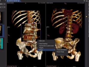
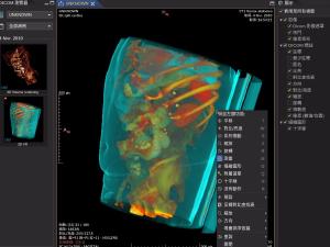
- Volume rendering with 3D presets
- Layouts for comparing series or studies
- Advanced series synchronization options
- Display cross-lines
- 3D cursor
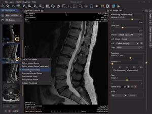
- Oblique Multi-planar Reconstruction (MPR)
- Maximum Intensity Projection
- Persistent magnifier glass
-
Measurement and annotation tools
- Length, area, and angle measurement
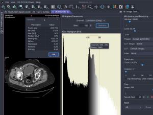
- Region statistics of pixels (Min, Max, Mean, StDev, Skewness, Kurtosis, Entropy)
- Histogram of modality values
- SUV measurement
-
Specific viewers
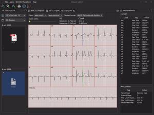
- DICOM ECG: display all the DICOM waveforms and allow to make some measurements
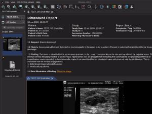
- DICOM SR: structured report viewer with hyperlinks to images and associated graphics
- DICOM AU: audio player (allow to export to WAV files)
-
Other tools
- Dicomizer module to convert standard images into DICOM
- Printing views to DICOM and system printers
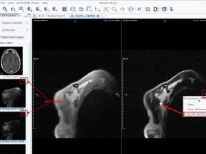
- Apply and Create DICOM Key Object Selection by selecting images with the star button
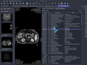
- Display and search into all DICOM attributes
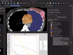
- DICOM RT tools for radiotherapy: display RT structure set, dose, and DVH chart
